
Research
Opening the book: separating twins without harming their brains
![[Twin A] and [Twin B] after their separation, holding hands with their father.](https://images.squarespace-cdn.com/content/v1/6ab50a3692cafe4288df60a8/3f33601c-6529-4ce0-b555-c91f9f49f026/Screenshot+2026-10-08+at+16.35.25.png)
Mercy and Goodness were born joined at the head, with their brains pressed closely together. A technique developed by our team, inspired by something as simple as opening a book, allowed surgeons to separate them without injuring either child's brain.
The challenge
When twins are joined at the head, their brains can sit so close together that there is almost no space between them. To separate the twins, surgeons have to work inside that narrow gap.
Until now, making room has usually meant holding the brains gently apart with instruments. But brain tissue is delicate, and even gentle, steady pressure can bruise it.
Mercy and Goodness were especially challenging since they were referred to Gemini after 1 year old, when the chances of a succesful separation drops more than 50%.
Like opening a book
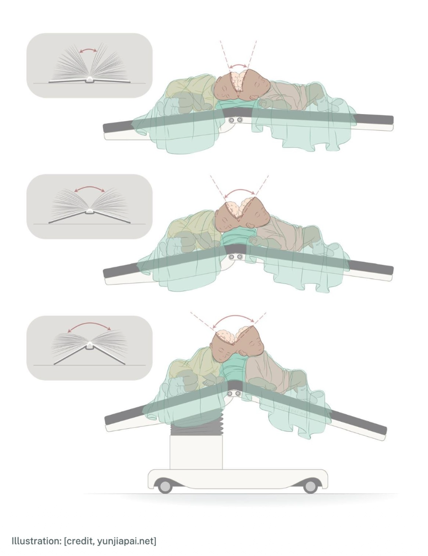
Instead of pushing the brains apart, our team let gravity do the work.
The twins lie on an operating table that can bend in the middle. The place where their heads are joined sits right above the hinge, like the spine of a book.
As the surgeons work, the two sides of the table are lowered a little at a time, like the covers of a book being opened.
With each step, the gap between the two brains widens on its own. The surgeons can see and work clearly, and nothing has to press on the brain.
Why it matters
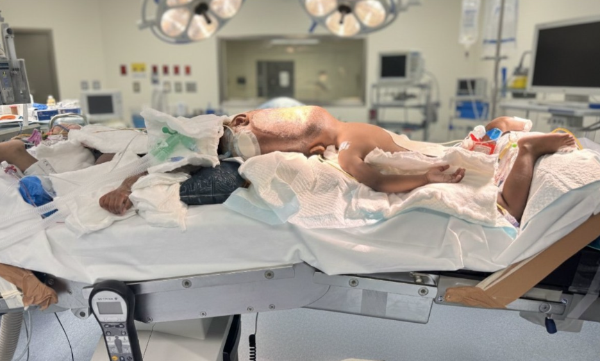
Because the space is created by the position of the children's bodies rather than by instruments, Mercy and Goodness were separated without any injury to either brain.
The idea came to Professor Jeelani the way many good ideas do: while he was doing something completely different. Reading a bedtime story to his son, he noticed that as the book opened wider, the pages fell apart on their own while the spine stayed put. Could a table that opened like a book let gravity gently separate two brains?
The difference it made
Today, Mercy and Goodness are thriving. They are walking on their own, exploring the world side by side, and growing into happy, curious children.
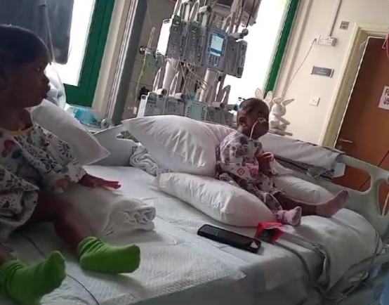
Sitting up on their own, side by side ater surgery
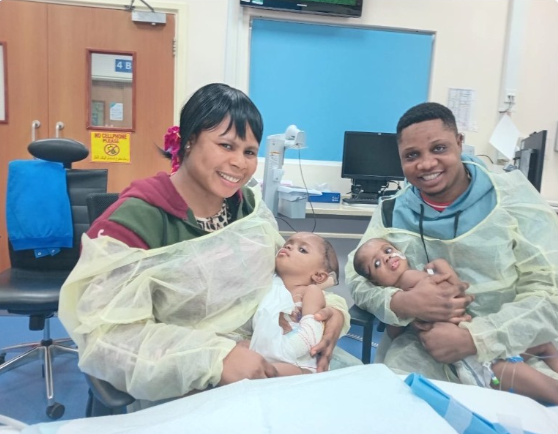
Held separately by their parents for the first time.
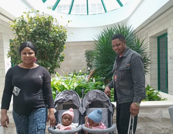
Out with mum and dad, each in their own pushchair.
Every advance starts with research
Ideas like this come from years of careful study by our surgeons, engineers and scientists. Your support helps us find safer ways to care for the children who need it most.
Made to measure: rebuilding a skull for each twin
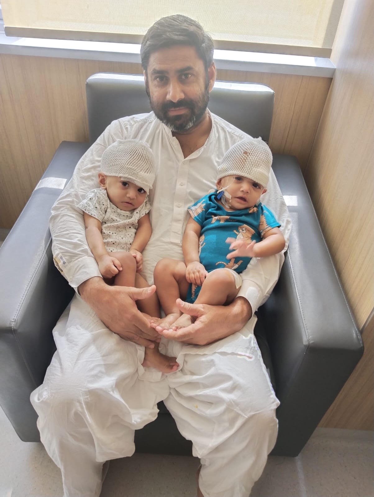
Mirha and Minal, the eighth set of twins separated by our team, were born joined at the head. After separation, each girl needed a new skull and enough skin to cover it. Our team designed both in our own lab, shaped for them alone.
The challenge
Separating twins who are joined at the head is only half the journey. Afterwards, each child is left with an opening in the skull where they were once connected, and it needs to be closed safely.
Standard skull implants are made for ordinary head shapes. Twins heads are shaped very differently, and each child's brain needs its own amount of room to grow. On top of that, twins never have enough skin to cover the wound once they are apart.
A new skull, designed in our lab
Instead of adapting an off-the-shelf implant, our team designs each reconstruction from scratch.
We start with the twins' own scans and build an exact 3D model of their heads on the computer.
A prediction model, developed through our research, works out how much space each twin will need, so the new skull fits the child and not just the gap.
The design is sent to an approved medical device manufacturer, who 3D-prints the skull pieces to our exact plan.
In surgery, each child receives a reconstruction made for them, unlike any other.
The two custom skull implants, one for each twin, tested on 3D-printed models of their heads.
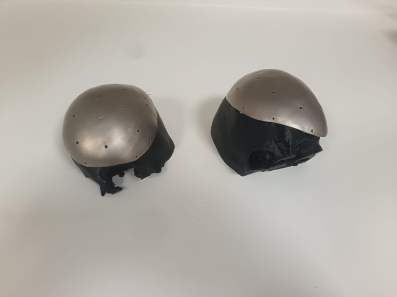
Growing the skin they need
To make sure there is enough skin to close each wound, we place skin expanders under the scalp months before separation. These are small balloons that are slowly filled over time, so the skin above them gently stretches and grows.
Here too, every detail is planned in our lab. We model the expanders to produce exactly the amount and shape of skin each twin will need for their reconstruction. The expansion took 2 months.
Why it matters
A skull and scalp designed for each child give them protection, a natural head shape, and room for their brain to keep growing as they do.
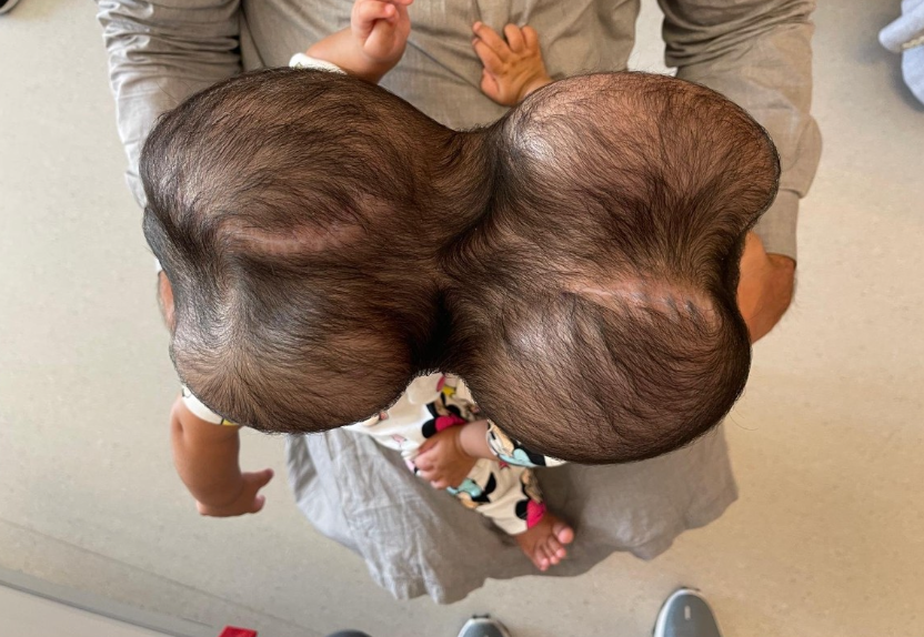
The difference it made
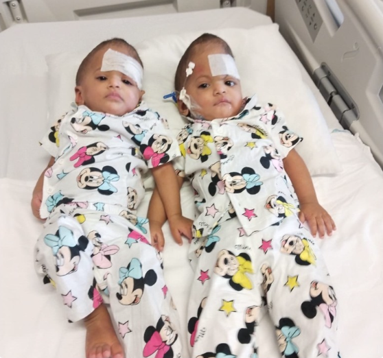
Side by side, now each in their own space. November 2025
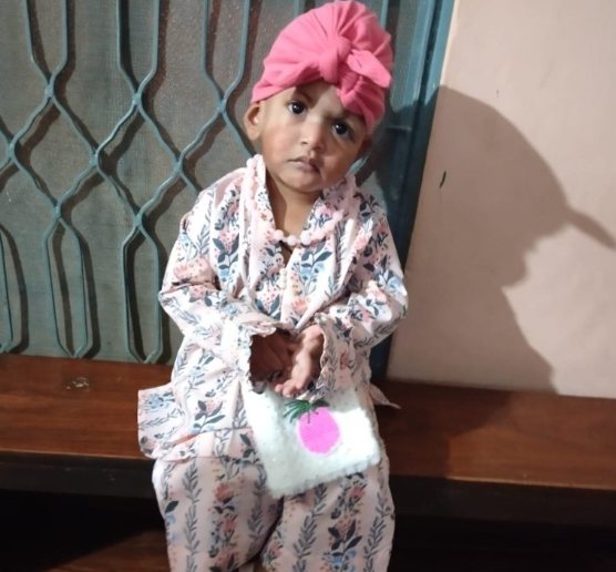
Mirha at home. July 2026
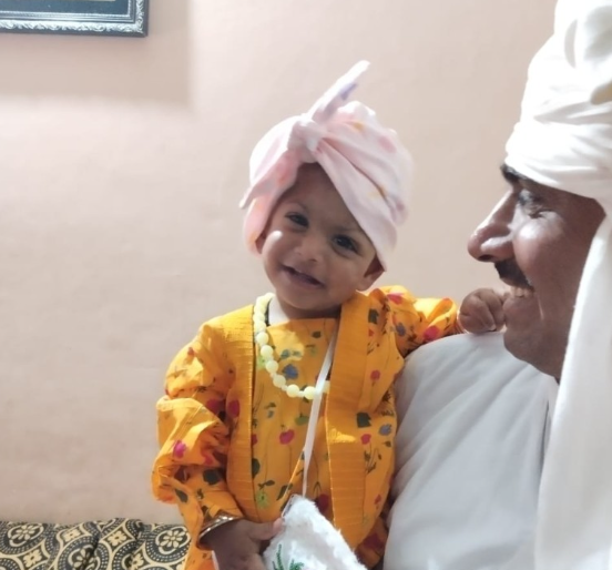
Minal at home. June 2026
Every advance starts with research
Ideas like this come from years of careful study by our surgeons, engineers and scientists. Your support helps us find safer ways to care for the children who need it most.
Practising every step before the real day
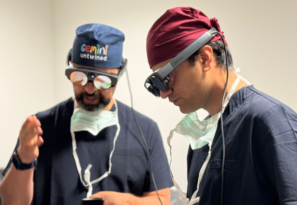
Before every separation, our surgeons rehearse the operation on life-size 3D-printed models and inside a virtual version of the twins' own anatomy. We have used this approach for our last three sets of twins: Arthur and Bernardo in Brazil, Mirha and Minal in Turkey, and Mercy and Goodness in Abu Dhabi.
No two sets of twins are the same
Every pair of twins joined at the head shares their skull, brain and blood vessels in a different way. There is no textbook that shows surgeons exactly what they will find.
During the operation, much of this anatomy is hidden from view. A surprise in theatre can be dangerous, so the more the team understands before they begin, the safer the children are.
A rehearsal for every operation
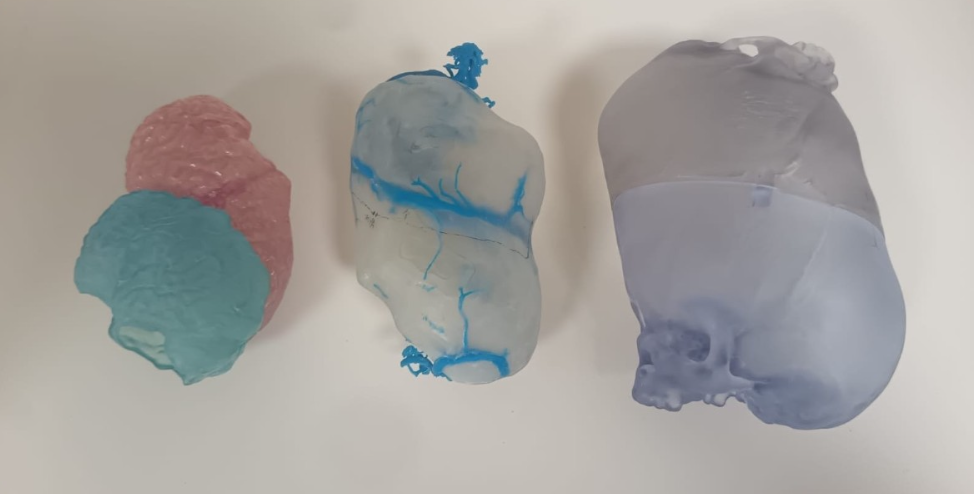
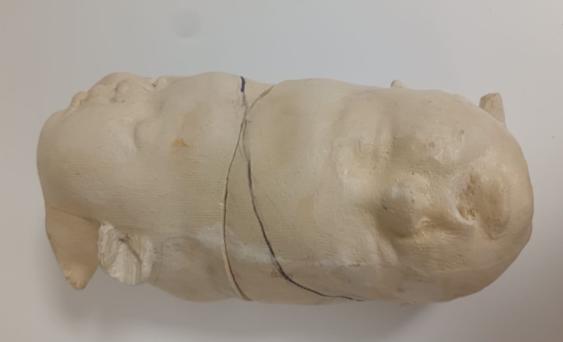
Using the twins' own scans, our lab builds an exact copy of their anatomy that the team can study, hold and practise on.
From the scans, we build a detailed 3D computer model of the twins' skulls, brains and blood vessels.
We 3D-print these parts life-size, in clear and coloured materials, so surgeons can hold them, turn them over and mark exactly where to work.
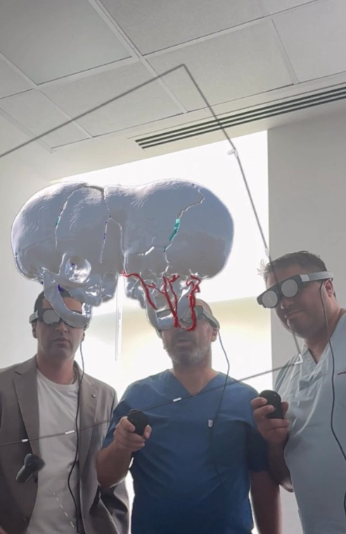
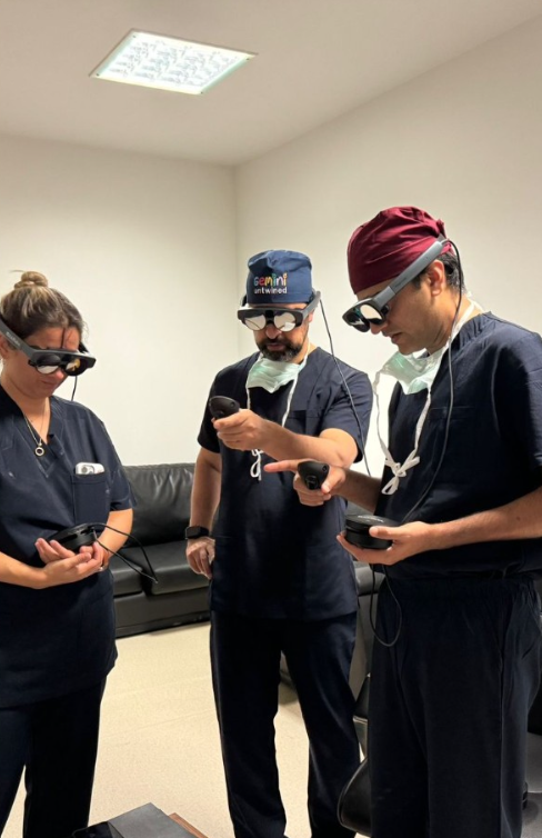
Wearing headsets, the whole team steps into a virtual version of the same anatomy. They can walk around it, look inside it and follow a single blood vessel from end to end.
Together, they rehearse each stage of the operation and agree the plan before the twins ever come into theatre.
Stepping inside the plan
In virtual reality, the twins' skulls and blood vessels float in the room, many times larger than life. Surgeons, engineers and specialists can stand around the same model, point to the same vessel and talk through every decision.
It means that when the real operation begins, the team has already seen what lies ahead.
Three families, one approach
Rehearsing in this way has become part of every separation we carry out.
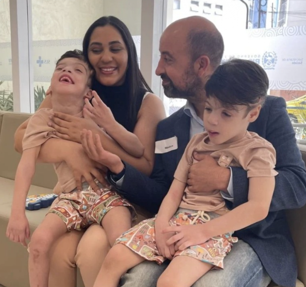
Arthur and Bernardo
Brazil, 2024. Very happy children enjoying life.
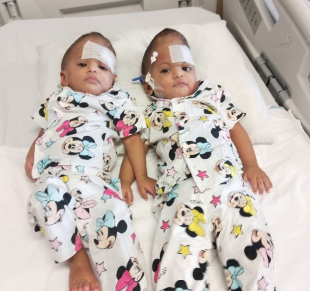
Mirha and Minal
Turkey, 2025. Living happy independent lives
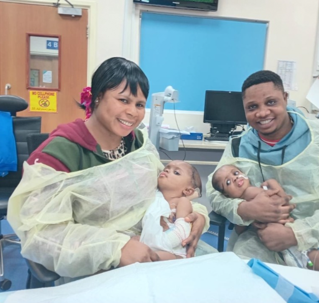
Mercy and Goodness
Abu Dhabi, 2026. Now walking on their own and two very happy children.
How You Can Help
We rely on the help from other people’s generosity. If you’re interested in helping please do follow the link below or fill in the form here. We’d be extremely grateful.